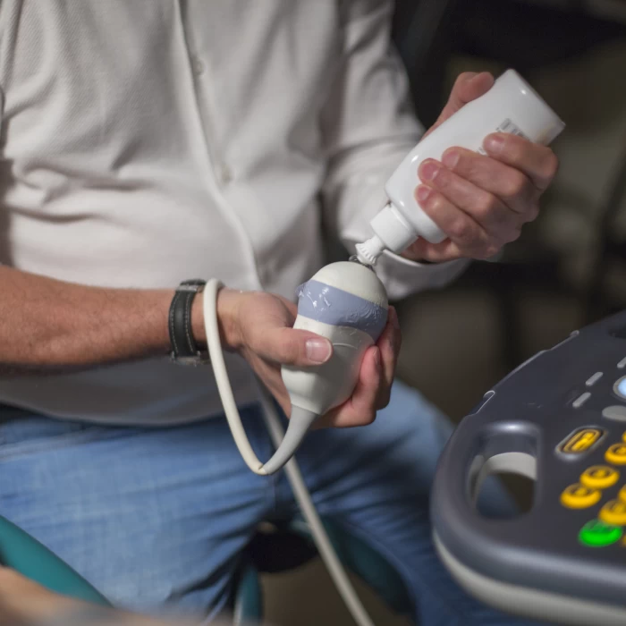
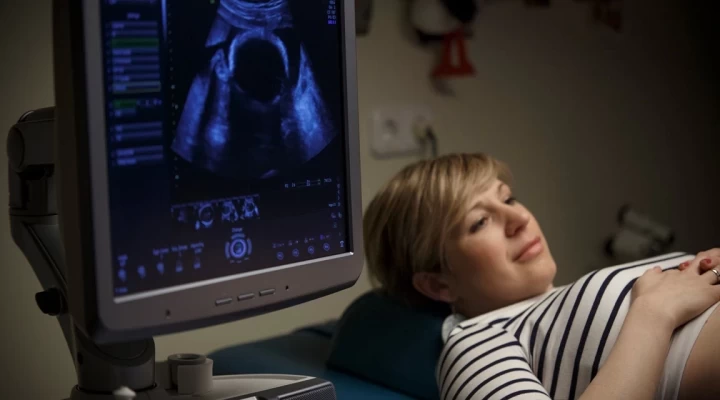
With these state-of-the-art ultrasound devices, it is possible to examine the fetal cardiovascular system within the womb. We can check the anatomy of the heart and measure the function of the heart valves, myocardium, flow and heart rate, so the majority of heart development disorders and arrhythmias can be detected when the baby is still a fetus. In addition, it is also possible to look for other organ and chromosomal abnormalities. The examination is performed through the abdominal wall, using an ultrasound probe specially developed for the examination of the heart. We can effectively reconstruct the heart from multiple intersection planes in 3D to ensure the majority of complex heart defects can be recognized.
When is this examination recommended?
- If a heart defect or arrhythmia is suspected during the obstetric screening examination, or if too much or too little amniotic fluid is found in the pericardium, fluid is measured in the chest cavity, abnormal flow is measured, thick neck folds and other organ development abnormalities are detected.
- Congenital heart problem or arrhythmia in the family history: in parents, previous child, or relatives
- The expectant mother has diabetes, connective tissue or autoimmune disease, if she has taken certain medicines that are harmful to the fetus during pregnancy (e.g. lithium, a medicine used to treat epilepsy), has had a viral infection
- Twin/multiple pregnancy
- The baby was conceived after artificial insemination
- Chromosome aberration is suspected (2/3 of babies with Down syndrome have a heart abnormality)
- Mother aged 37 or above
Our related doctors
Any questions before booking an appointment?
If you are unsure which doctor to see or what examination you require, we are here to help!
Simply request a free callback from one of our colleagues, who will help you find the right specialist based on your specific issue.


Reviews