
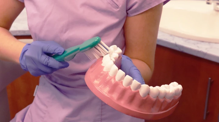
A thorough oral examination provides a great deal of information on its own, but accurate diagnosis and the planning of safe, long-term successful treatments require radiographic imaging. Today’s technology allows us to produce highly detailed images with minimal radiation exposure—whether in two-dimensional or three-dimensional form. But when is a panoramic X-ray sufficient, and when does a 3D CT scan, also known as CBCT, become necessary?
Two commonly used dental imaging methods are the 2D panoramic X-ray and the 3D CT. At first glance, they serve a similar purpose—mapping the condition of the oral cavity—but their operation and the depth of information they provide differ significantly. A panoramic X-ray produces a flat, two-dimensional image showing the entire dentition, the jaws, and the temporomandibular joints at once. This type of image is an excellent starting point, as it quickly and comprehensively reveals potential issues such as missing teeth, cavities, inflammatory lesions, or even hidden abnormalities.
However, it is important to understand that panoramic X-rays have inherent limitations. Since three-dimensional anatomical structures are projected onto a single plane, certain details may overlap, become distorted, or appear less sharp. This means that while it provides an excellent general overview, it may not offer sufficient detail for precise treatment planning in some cases.
This is where 3D CT, or CBCT (Cone Beam Computed Tomography), comes into play. This technology allows the examined area to be analyzed in three dimensions, layer by layer. The image can essentially be “sliced,” enabling the clinician to examine the region from every angle. This not only provides a more detailed view but also allows for precise measurements, which are crucial for many procedures.
One of the greatest advantages of CBCT is that it provides distortion-free images in true scale. Bone thickness and density become clearly visible, as do the course of nerves, the exact number and direction of root canals, and even hidden abnormalities that might go unnoticed on a 2D X-ray. This level of detail is particularly important when treatment planning requires millimeter-level precision.
A prime example is implant planning. A panoramic X-ray clearly shows where a tooth is missing and allows for an approximate estimation of the available bone height. However, bone width—which is equally important for the stable placement of an implant—cannot be accurately assessed. It’s like looking at a mountain from the side: you can see its height, but you have no idea how wide it is.
In contrast, a 3D CT scan allows precise measurement of bone height, width, and density. It also clearly shows the course of nerves, especially in the lower jaw, where proximity to the nerve is a critical factor. If an implant is placed too close to a nerve, it can cause unpleasant complications such as numbness or persistent pain. For this reason, CBCT has become almost a standard requirement for safe implant planning.
Similarly, 3D imaging plays an important role in planning the removal of wisdom teeth. While a panoramic X-ray can show the angle of the tooth and indicate that a nerve runs nearby, it does not provide an exact picture of their spatial relationship. This uncertainty can pose risks during surgery.
CBCT, on the other hand, visualizes the relationship between the tooth and the nerve with millimeter precision. The clinician can clearly see whether the nerve runs beside, below, or even between the roots. This information significantly influences the surgical technique and, in some cases, even the decision of whether the procedure is necessary. A well-planned intervention is not only safer but also results in faster healing and less discomfort for the patient.
To illustrate the difference with an everyday analogy: a panoramic X-ray is like a map. It shows direction and helps with orientation but does not provide every detail. CBCT, in contrast, is like using a GPS combined with a three-dimensional model—you can clearly see the destination, the route, and any potential obstacles.
It is important to emphasize, however, that a 3D CT scan is not necessary in every case. For routine check-ups or basic examinations, a panoramic X-ray is often perfectly sufficient. The goal is always to achieve an accurate diagnosis with the least possible intervention and the greatest comfort for the patient.
At the same time, there are situations where CBCT is essential to ensure maximum safety and precision. In such cases, it is not an “extra” service but part of responsible, modern care. At our private clinic in Buda, our advanced CBCT equipment allows diagnosis and treatment planning to be carried out at the highest professional level, minimizing risks and maximizing long-term success.
Choosing the appropriate imaging method is always an individual decision, determined by the treating dentist based on the patient’s current condition and the planned procedure. What is certain, however, is that modern diagnostics not only provide greater accuracy but also ensure safer dental care.
- dietetics25
- pediatrics24
- diet18
- RMC Dental Center15
- internal medicine11
- dentistry9
- Dr Kinga Jókay8
- pediatric dentistry8
- sport7
- zsuzsanna kókai6
- coronavirus6
- screening6
- covid6
- urology6
- obstetrics gynecology6
- prevention5
- cardiology5
- allergy5
- psychology5
- dental hygiene5
- infection4
- gastroenterology4
- menopause4
- screenings3
- Kinga Jókay M.D3
- Flu shot3
- Flu3
- women's screening3
- Dr Nóra Gullai2
- infectious disase medicine2
- Ear-Nose-Throat2
- Same Day Surgery Center2
- heat stroke2
- endocrinology2
- Dr Tímea Baló2
- allergen immunotherapy2
- Adrienne Nagy M.D.2
- Fetal Medicine Center2
- pediatric urology2
- flu season2
- meningitis2
- dermatology2
- gynecologist2
- Ophthalmologist2
- Headache Clinic2
- Dr Eszter Bodnár2
- headache2
- menopause program2
- invisalign2
- diabetology2
- cardiovascular disease1
- diabetes1
- hydration1
- hematology1
- iron1
- Imre Bodó M.D.1
- nutritional disorder1
- lyme disease1
- gynecologic oncology surgery1
- first trimester screening1
Our related doctors

Any questions before booking an appointment?
If you are unsure which doctor to see or what examination you require, we are here to help!
Simply request a free callback from one of our colleagues, who will help you find the right specialist based on your specific issue.